CM - Port Wine
What is a Capillary Malformation?
Capillary Malformations (CMs) are abnormal growth of capillaries, the blood vessels that deliver blood to tissues by receiving blood from arteries and draining into veins. Capillary malformations, or port-wine stain, are most commonly present at birth and appear as flat, irregularly shaped, reddish purple discolorations of the skin. They are often located on the head and neck and typically on one side. There are multiple different types of capillary malformations, but two of the more notable types present at birth include port wine stains and salmon patches. Salmon patches differ from port wine stains in that they tend to be smaller in size and frequently regress with age. The majority of this page will focus on port wine stains as these do not regress and thus require treatment.
What causes capillary malformations?
Capillary malformations can be associated with maternal age greater than 35, and are also associated with several underlying medical conditions, namely Sturge Weber, Proteus Syndrome, Roberts Syndrome, Wyburn-Mason Syndrome, and Cobb Syndrome. However, the majority of cases are sporadic, meaning there is no known familial history. They occur equally in both males and females in about 0.1-0.3% of births. A thorough workup is recommended by a vascular anomaly specialist after identification.
The underlying cause of capillary malformations are not well understood but there are a few theories: One theory suggests that abnormal signaling in the form of too much growth stimulation from a molecule called Vascular Endothelial Growth Factor (VEGF) paired with an abnormally high number of receptors for VEGF leads to overgrowth of capillaries. This theory is supported by genetic studies which suggest that mutations in a gene that regulates VEGF on chromosome 5 is often mutated in individuals with port wine stains. Another theory holds that abnormal formation or function of the nervous system near the port wine stain is responsible for the capillary malformation. This theory is supported by the fact that the capillaries in CMs are not as responsive to certain nervous system signals (catecholamines) that work to constrict the capillaries, suggesting this may lead to their subsequent dilation and overgrowth. Additional evidence for this theory comes from the fact that port wine stains often take the shapes similar to patterns of the underlying neural innervation in that area (dermatomes).
Clinical Presentation and Diagnosis
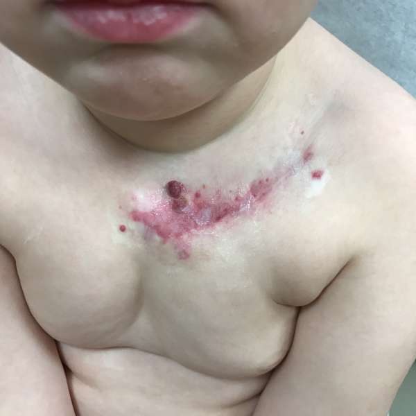
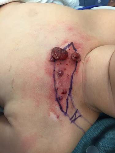
At birth Capillary malformations appear as reddish purple patches on the skin, often resembling a streak of spilled wine, hence the name “port wine stain.” Histologically, or underneath the microscope, capillary malformations initially appear as tiny thin walled superficial (close to the surface of the skin) vessels. Over time, these vessels dilate and thicken due an increase in reticulin, a structural molecule, found in the basement membrane, or supportive floor of the vessels. With time, and possibly related to hormonal changes, vascular growths (blebs) can appear, causing bleeding and pain.
Diagnosis of CMs is mainly done by their distinctive appearance however a tissue sample can be taken for confirmation if necessary.
Complications
The main complications come from aesthetic issues associated with the capillary malformations. CMs grow proportionately as a child gets older and the majority of them begin to show a bumpy nodular pattern of growth by the time the patient reaches their 50s. Further, given that many are located on the head and neck this can be very challenging for a child and for this reason many parents seek treatment for capillary malformations.
Additionally, capillary malformations can lead to an overgrowth or enlargement of the underlying soft tissue and bone, leading to an asymmetric appearance that can also be aesthetically displeasing. This overgrowth is thought to be due to the additional blood supply to the underlying tissues and bones from the additional capillaries there.
Treatment
Unfortunately treating capillary malformations can be a challenging process. Current treatments include the use of pulsed dye laser and surgical resection. Additional studies involving local use of certain anti-cancer and immune system suppressing drugs are also currently underway.
Pulsed Dye Laser
The safest and most efficacious method entails the use of pulsed dye laser. This entails setting the laser to a specific wavelength (585nm) and turning the laser on and off in a pulsatile fashion. The underlying success of this technique is due to the fact that CMs contain a high quantity of a specific cellular component called chromophore oxyhemoglobin, which is targeted by the specific wavelength used in the laser. Destruction of this component leads to subsequent destruction of the capillaries and thus fading of the CM.
The pulsed dye laser is often used consecutively over multiple sessions until there appears to be no more improvement in the CM. The main complication of this treatment is scarring or irregular pigmentation of the skin in the treated area. Additionally, while pulsed dye laser is the most effective treatment, studies have shown that up to 20% of cases may be resistant to treatment with pulsed dye laser, meaning that the CM does not lighten with use. In these cases surgical resection may be indicated. Several sessions are often necessary, usually around 5-7, to effectively treat the problem. Numbing cream can be applied 1-2 hrs before treatment to help reduce some of the discomfort. The actual process takes only a few minutes and the aftercare is similar to that of a sunburn. The PDL laser treatment is most often covered by most medical insurances.
Surgical Resection
While not the first line choice of treatment, surgical resection of CMs is effective. It entails the removal of the CM as well as any other vascular malformations that may be associated with the area. Surgery may often be combined with laser to completely remove capillary malformations, particularly in anatomically sensitive areas. Surgery is also most effective for removing the bumps (blebs) often associated with capillary malformations. The laser can only penetrate around 1mm, so surgery is more effective for the areas that are raised.
Experimental Treatments:
Prior studies have indicated that the use of Sirolimus, a molecule that prevents inflammation by inhibiting the communications between inflammatory cells, was not an effective adjunct to pulsed dye laser and failed to improve outcomes.
A current clinical trial is underway for the use of bleomycin sclerotherapy with electroporation. Bleomycin is a cancer drug that damages cellular DNA and is used to induce apoptosis (or cell death). When injected into larger vascular malformations in a process known as sclerotherapy, it causes the death of the vascular cells and subsequent decrease in blood flow through the area. In the past, bleomycin has not been used for CMs because it has been challenging to deliver the drug to the appropriate cells given the small size of the capillaries, as capillaries are the smallest of blood vessels. A current clinical trial underway examines the use of electroporation- a process that essentially uses electricity to zap the cells and make them more permeable and increase the probability of delivering bleomycin to the cells.