What are Arteriovenous Malformations?
During embryogenesis when the blood vessels are forming in the fetus, arteries and veins are formed first and then the capillaries (which are supposed to deliver the blood to the tissue) are formed second. Arteriovenous Malformations (AVMs) result when something goes wrong with this process resulting in a direct connection between the arteries and veins without the formation of a true bed of capillaries between them.
Normally, capillary beds are responsible for providing resistance to flow so that blood cannot rush from artery to vein. As AVMs do not have capillary beds, there is a high speed flow of blood from arteries and veins, and the veins experience a resultant higher level of pressure than they normally endure, leading to changes of venous appearance and complications (both are described further below)
AVMs are composed of what are called “feeding arteries” and “draining veins” and a space of tangled arteries and veins between them called the nidus. The exact nature and appearance of the nidus depends on where things went wrong exactly during emrbyogenesis. AVMs that result from an error in early embryogenesis are called Extratruncular AVMs and those that occurred later are called Truncular AVMs.
Extratruncular AVMs are composed of very early embryonic tissue called “mesenchyme” and have a well-defined nidus (source). Truncular AVMs are composed of cells during a later stage of development and resultantly do not have mesenchymal cells; they have a poorly defined nidus. Both types of AVMs are progressive and grow over time.
AVMs are mainly congenital vascular malformations (appear at birth) but they may also develop sporadically later in life if a patient has an underlying illness leading to a genetic predisposition such as Cerebrofacial Arteriovenous Metemeric Syndrome, Capillary Malformation Arteriovenous Malformation Syndrome, or Hereditary Hemorrhagic Telangiectasia (HHT). About 70% of AVMs occur in the head and neck region, with many of them occurring intracranially (around the brain).
Complications if Untreated
Complications of AVMs are due to both their inherent structure, as well as their proliferative nature:
- Aneurysm- Given the fast flow and direct connection between veins and arteries, veins are under pressure that they are not accustomed to and often undergo aneurysms. This can lead to bulging and potential rupture of veins, causing bleeding. If this occurs in the brain this can result in neurological deficits. In other areas of the body, the bleeding can lead to a decrease in blood delivery to surrounding tissues resulting in tissue damage.
- Facial Asymmetry- If the AVM involves bones of the face, it can result in hypertrophy (or increased growth) of the bone due to the extra nutrients delivered from the AVM. This can result in severe facial asymmetry and a dysmorphia.
- Cardiac Failure - Overtime as the AVM grows, it can lead to heart failure if too much blood is shunted directly from the arteries in the AVM to the veins. This returns blood to the right side of the heart too quickly, creating too much pressure and overwhelming the right side of the heart. This puts undue strain on the heart and can cause heart failure.
Clinical Presentation and Diagnosis:
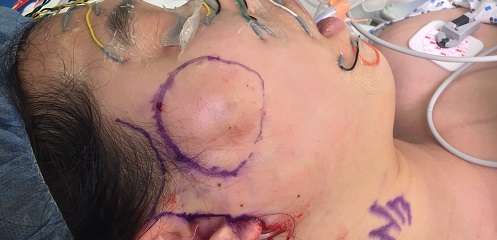
AVMs that are close to the skin may present at birth as poorly defined flat red rashes, however some AVMs may not present until puberty, and others do not present at all and are incidentally discovered later in life. Those that are close to the skin progress over time to become warm, pulsating red to purple colored masses that are difficult to compress. A pulse may be felt when touching these AVMs and audible whooshing of blood may be heard. In some cases, AVMs may appear later in life as a result of proliferation in response to a stimulus such as trauma, surgery, hormonal changes, or pregnancy.
Ultrasound with Doppler is used to confirm the presence of AVM as it shows the rapid movement of blood directly from artery to vein without an intermediate capillary bed. After ultrasound, MRI is often done to assess the extent, severity, and nature of the AVM with regards to surrounding tissue. This information is useful in informing treatment. Other less commonly used methods of diagnosis include CT scan with contrast, CT scan with three dimensional reconstruction, and whole body blood pool scintography (a method which entails labeling red blood cells with a radiopharmaceutical substance that can be captured with a specific imaging device).
A tissue biopsy can be performed during the course of diagnosis and treatment, however it has limited utility as the results are dependent on the sample, and not all samples will contain all the elements of the AVM necessary for concrete diagnosis. Biopsy may reveal a mix of large arteries and veins as well as some small vessels in a fibrous myxoid (or mucoid looking) background. Distorted arteries can be seen directly connecting to veins, and veins show signs of withstanding high speed high pressure flow. Typically veins have a thin wall and very little muscle surrounding them however veins in AVMs show thickening of the muscles and the fibrous structure in the walls in response to high pressure and high flow.
Treatment:
The goal of treatment is to either remove or destroy the entire AVM or the entire nidus (source of the problem). This is because in the past, AVMs, particularly the extra-truncular type, treated without complete nidus resection have been shown to frequently recur. Current theory holds that the mesenchymal cells of the extratruncular AVMs lie in the nidus and regenerate the AVM when incomplete resection occurs. Removal or destruction of the nidus is thus important to prevent AVMs from returning after excision or ablation.
Exact treatment depends on the size, location, and extensive nature of the AVM.
- Small AVMs: Very small AVMs that are well defined (not significantly invading surrounding tissues) can be treated directly with surgical resection. Stealth approaches using facial shadows can be applied to minimize facial scarring.
- Large AVMs: Poorly defined and large AVMs must be treated with embolization prior to surgery. Embolization entails injecting a substance into the nidus of the AVM to induce the formation of a blood clot. Common substances used for embolization of AVMs include ethanol, glue, and coil therapy. Ethanol both damages the AVM directly as well as induces a clot, making it more effective than glue and coil therapy which only induce a clot. However, this efficacy comes at a higher risk of damage to the tissue surrounding the AVM. When done prior to surgery, embolization helps decrease the size of the AVM by decreasing the blood flow there, making it easier to resect and decreasing the risk of severe blood loss during surgery. In cases where AVMs are unable to be completely resected during one surgery, multiple rounds of embolization and surgical resection may occur.
- Intracranial AVMs: When AVMs are intracranial, a special technique called radiosurgery is sometimes used, which entails delivering a high dose level of ionizing radiation directly to the nidus. Radiotherapy has been shown to be highly effective when given at high doses, resulting in full AVM destruction in up to 90% of cases, however complications of high dose radiosurgery may include neural damage to surrounding brain tissue.
When to Operate:
Given the severity of the complications associated with AVMs, it is generally recommended that all AVMs be operated upon given that patient is medically stable for operation. If the risks of surgery outweigh the benefits as would happen in a patient with poor underlying health, the AVM can be monitored. Combination therapies are often used to embolize and then remove AVMs to minimize complications.